Venovenous extracorporeal membrane oxygenation (VV ECMO) is a life-support technique used to provide respiratory support to patients with severe respiratory failure. It oxygenates the blood and removes carbon dioxide externally, allowing the lungs to rest and heal. Below are the key configurations and components of VV ECMO:
1. Cannulation Configurations
Cannulation refers to how the tubes (cannulas) are placed to access the patient’s blood vessels. Common VV ECMO configurations include:
- Femoral-Jugular Configuration:
- Drainage Cannula: Placed in the femoral vein (large vein in the groin) to deoxygenated blood from the body.
- Return Cannula: Placed in the internal jugular vein (neck) to return oxygenated blood to the right atrium.
- Advantages: Good for larger patients or when high flow rates are needed.
- Disadvantages: Risk of recirculation (oxygenated blood being pulled back into the ECMO circuit).
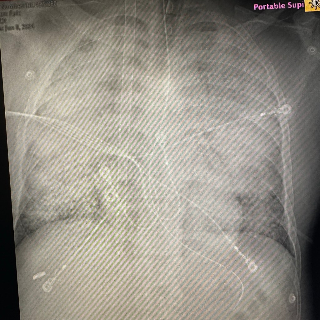
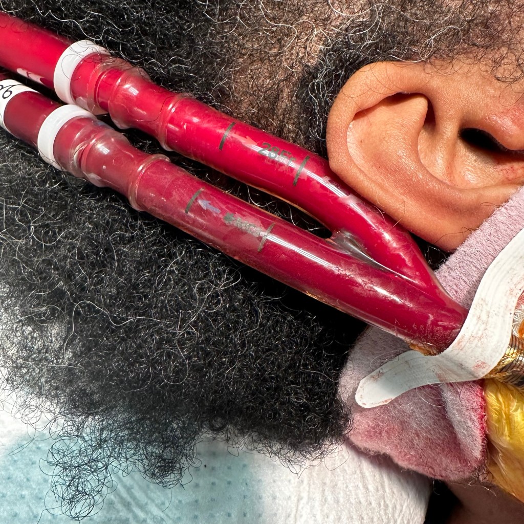
- Dual-Lumen Cannula (Avalon or Crescent Cannula):
- A single cannula placed in the internal jugular vein with two lumens: one for drainage and one for return.
- Advantages: Reduced recirculation, easier patient mobility, and fewer access sites.
- Disadvantages: Technically challenging to place, risk of malposition, and limited flow rates.
- Femoral-Femoral Configuration:
- Both drainage and return cannulas are placed in the femoral veins.
- Advantages: Simpler cannulation.
- Disadvantages: High risk of recirculation and limited oxygenation efficiency.
2. ECMO Circuit Components
The ECMO circuit consists of several key components:
- Cannulas: Tubes inserted into the veins to access blood.
- Pump: Provides continuous blood flow through the circuit (usually a centrifugal pump).
- Oxygenator: Acts as an artificial lung, oxygenating the blood and removing carbon dioxide.
- Heater-Cooler Unit: Maintains blood temperature.
- Tubing: Connects all components and carries blood.
- Monitoring Systems: Track flow rates, pressures, and oxygen levels.
3. Blood Flow and Recirculation
- Flow Rates: Typically range from 2 to 6 liters per minute, depending on patient size and needs.
- Recirculation: Occurs when oxygenated blood from the return cannula is drawn back into the drainage cannula, reducing efficiency. Proper cannula placement and configuration minimize this.
4. Indications for VV ECMO
- Severe acute respiratory distress syndrome (ARDS).
- Refractory hypoxemia (low oxygen levels despite maximal ventilator support).
- Bridge to lung transplantation.
- Severe respiratory failure due to pneumonia, trauma, or other causes.
5. Advantages of VV ECMO
- Provides full respiratory support.
- Allows lung-protective ventilation strategies.
- Reduces ventilator-induced lung injury.
6. Challenges and Risks
- Complications: Bleeding, infection, clotting, and cannula-related issues.
- Recirculation: Reduces oxygenation efficiency.
- Hemodynamic Instability: Requires careful monitoring.
- Resource-Intensive: Requires a specialized team and equipment.
7. Monitoring and Management
- Regular blood gas analysis to assess oxygenation and carbon dioxide removal.
- Monitoring of circuit pressures and flow rates.
- Anticoagulation management to prevent clotting in the circuit.
VV ECMO is a complex but life-saving therapy for patients with severe respiratory failure. Proper configuration, monitoring, and management are critical for successful outcomes.


Leave a comment